Although imperforate hymen is the most common congenital anomaly of the female genital tract, it remains an uncommon diagnosis in the emergency department. Patients often present to emergency physicians with non-specific symptoms such as lower abdominal pain, urinary retention, constipation or back pain, making the diagnosis easy to miss. Ultrasound can rapidly identify the condition at the bedside and expedite definitive management
Case
- An 11 -year-old girl presented to the emergency department with worsening lower abdominal pain and difficulty passing urine.
- On examination she has a distended tender abdomen.
- She was initially thought to be in urinary retention. She was catheterised but had very little urine in her bladder.
- A point of care ultrasound was performed after catheterisation.
POCUS Findings
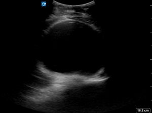
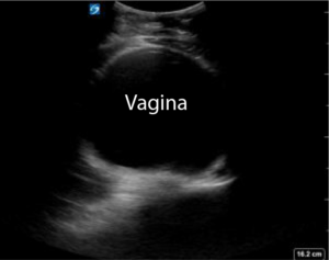
What do you make of the ultrasound findings . The first image was taken with the probe in the longitudinal plane in the pelvis. The second scan was taken in the transverse plane.
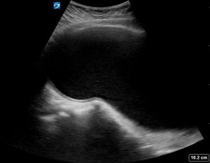
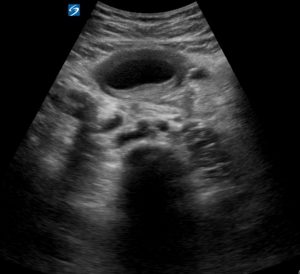
The transabdominal ultrasound demonstrated:
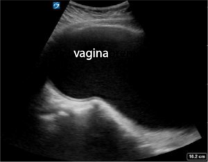
- A markedly distended vagina filled with echogenic fluid (hematocolpos)
- A large midline cystic pelvic mass located posterior to the bladder
- Compression of the bladder by the distended vagina
The bladder can be distinguished from the vagina by it’s thicker walls.
Longitudinal view
The vagina is grossly distended and filled with fluid.
Transverse view
The bladder can be distinguished from the vagina by it’s thicker wall.
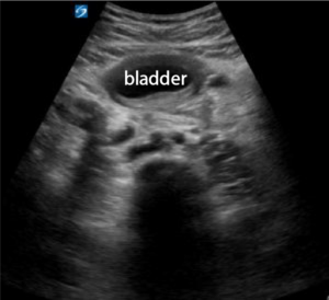
Transverse view of the vagina
What is an Imperforate Hymen?
How does an imperforate hymen typically present clinically?
What would you find on POCUS?
What is the management ?
POCUS Pearls
- Primary amenorrhoea with cyclical abdominal pain is imperforate hymen until proven otherwise.
- A large midline cystic pelvic mass behind the bladder should prompt careful evaluation of the vagina and uterus.
- Bedside ultrasound allows rapid diagnosis and may prevent unnecessary CT imaging.
- Always perform a focused external genital examination when the history suggests an obstructive genital tract anomaly.
Updated August 2026


Leave A Comment