Abdominal pain is one of the most common presentations to the emergency department. While most children have benign causes of pain, clinicians must always remain alert for conditions where early diagnosis can dramatically change the outcome.
One such condition is intussusception.
Although it most commonly occurs between 6 months and 3 years of age, it can also present in older children, where a pathological lead point becomes more likely. Ultrasound is the imaging modality of choice, and point-of-care ultrasound (POCUS) allows emergency physicians to rapidly identify this surgical emergency at the bedside
A 6 year old girl presents to the Emergency department with dysuria and abdominal pain. The symptoms have been intermittent until the morning of the presentation when the symptoms were constant and also associated with a left sided limp. She is unwilling to weight bear on her left leg. She had not had a bowel motion since the night before. She has had no vomiting. On examination there is full painless passive movement of the left and right hips. She is tender in the left iliac fossa. No rebound tenderness or guarding. Urine microscopy shows no white, red cells or organisms. An abdominal xray is performed
Xray shows faecal matter within the caecum, ascending colon, sigmoid colon and rectum with paucity of gas within the descending colon. No pneumoperitoneum
POCUS of the bowel in the area of pain revealed the following:
Transverse View:
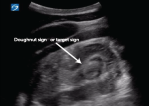
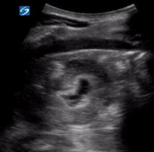
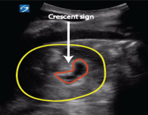
The transverse scan demonstrated the classic “target sign” (also known as the doughnut sign), consisting of multiple concentric hypoechoic and hyperechoic rings representing bowel telescoped within bowel.
Longitudinal View
The longitudinal scan demonstrated the characteristic “sandwich sign” or “pseudokidney sign”, another hallmark appearance of intussusception.
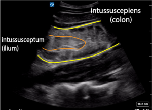
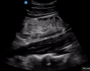
These sonographic findings confirmed the diagnosis of ileocolic intussusception.
Shortly after the ultrasound the patient vomited a large amount of green vomit followed by ongoing smaller clear vomits.
What is Intussusception?
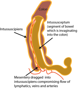
Intussusception occurs when one segment of bowel telescopes into the adjacent distal bowel.
This leads to:
- bowel obstruction
- venous congestion
- bowel wall oedema
- impaired arterial blood flow
- bowel ischaemia if untreated
The majority of paediatric cases are ileocolic and are idiopathic, often occurring after viral illnesses due to hypertrophied Peyer’s patches acting as a lead point. In children older than five years, however, pathological lead points such as Meckel diverticulum, polyps, lymphoma, or duplication cysts become increasingly important considerations.
Clinical Presentation
The classic triad of:
- intermittent colicky abdominal pain
- vomiting
- “red currant jelly” stool
is actually present in only a minority of patients.
More commonly, children present with:
- episodic severe pain
- crying with legs drawn to the chest
- vomiting
- pallor
- lethargy
- periods of complete normality between painful episodes
Some children may present predominantly with lethargy, making diagnosis challenging.
Ultrasound Tips
POCUS is ideally suited to diagnosing intussusception because the examination is quick, non-invasive and highly accurate in experienced hands.
A systematic approach includes:
- Start in the right lower quadrant.
- Sweep slowly across the colon.
- Identify the ascending colon and caecum.
- Follow the bowel proximally and distally.
- Scan in both transverse and longitudinal planes.
Look for:
✅ Target (doughnut) sign
✅ Sandwich sign
✅ Pseudokidney sign
Also assess for:
- free fluid
- bowel wall thickness
- bowel perfusion (if Doppler available)
- enlarged mesenteric lymph nodes
Experienced emergency physicians performing POCUS have reported sensitivities around 95–96% and specificities greater than 90% for ileocolic intussusception.
Management
Once diagnosed:
- Keep the child nil by mouth.
- Establish intravenous access.
- Begin fluid resuscitation if required.
- Provide adequate analgesia.
- Notify paediatric surgery early.
- Arrange urgent radiology for therapeutic air or contrast enema if appropriate.
Air or hydrostatic enema reduction is successful in approximately 80–90% of uncomplicated ileocolic intussusceptions. Surgery is required when non-operative reduction fails or if perforation or peritonitis is suspected.
Learning Points
- Consider intussusception in any child with intermittent severe abdominal pain and vomiting.
- The classic triad is uncommon—absence of bloody stool does not exclude the diagnosis.
- Bedside ultrasound allows rapid diagnosis before bowel ischaemia develops.
- The target sign and sandwich sign are the classic sonographic appearances.
- In children over five years, always consider an underlying pathological lead point.
Take-Home Message
Point-of-care ultrasound has become one of the most valuable bedside tools in paediatric emergency medicine. In children presenting with intermittent abdominal pain, recognising the characteristic ultrasound appearances of intussusception can dramatically shorten the time to diagnosis, expedite definitive treatment, and prevent bowel ischaemia.
References
- Emerg Med Australas. 2007 Feb;19(1):45-50. Paediatric intussusception: epidemiology and outcome. Blanch AJ1, Perel SB, Acworth JP
- N Z Med J. 2010 Oct 15;123(1324):32-40. Ten-year review of intussusception at Starship Hospital: 1998-2007. Kodikara H1, Lynch A, Morreau P, Vogel S
- Acta Chir Belg. 2015 Sep-Oct;115(5):327-33. Intussusception in Children: A Clinical Review. Charles T1, Penninga L, Reurings JC, Berry MC.
- https://www.uptodate.com/contents/intussusception-in-children
- Journal of Pediatric Surgery Case Reports Volume 2, Issue 5, May 2014, Pages 228-23 open access Reports Left sided colo-colic intussusception in a child with acute lymphoblastic leukemia: A case report and systematic review of the literature
Excellent images