| Image optimisation | Machine Choose correct probe - High frequency – more superficial, linear probe
- Lower frequency – depth curvilinear probe. Sector probe
Machine adjustment (Don’t Forget The Giant Family Dog) - Depth
- Frequency
- TGC
- Gain
- Focus
- Dynamic range
Patient - Position (left lateral/sitting/standing, supine)
- Breathing manoeuvres
- Operator
- Firm pressure with probe to improve contact
- Adequate gel to improve contact of probe with skin
- If gas present in abdominal exam – graded pressure or “bouncing” of probe to displace gas
|
| Image artifacts | - Acoustic enhancement
- Acoustic shadowing
- Reverberation artifacts (due to gas or air or between probe and underlying tissue,)
- Mirror image (reflection of soundwaves)
- Edge artifacts (refraction of sound waves)
- Beam width (side lobe artifacts)
- Artifacts due to different propagation speeds
- Twinkle artifact (renal stones) – cause unknown
|
| FAST Fluid in abdomen | RUQ- Fluid in morrison’s pouch – need to fan through
- Fluid at tip of liver
- Fluid between diaphragm and liver
- Fluid at inferior pole of kidney
- Fluid in costophrenic angle
- Fluid in right paracolic gutter
LUQ - Fluid between spleen and diaphragm
- Fluid between kidney and spleen
- Fluid in left paracolic gutter
Pelvis - Fluid in retrovesicular space
- Fluid in pouch of Douglas
- Fluid between loops of bowel
|
| DVT | - Lack of compressibility of vein
- Absence of colour flow
- Pulse Doppler – lack of augmentation if complete occlusion by thrombus
- Lack of respiratory phasicity on colour flow suggests proximal thrombus (eg as in May Thurner Syndrome)
|
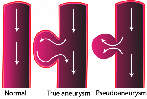
| AAA | - Aneurysmal if > 3cm
- Scan in long and in transverse axis
Features suggestive of rupture (rarely seen) - Periaortic haematoma
- Retroperitoneal haematoma
- Intraperitoneal fluid or haematoma
- Colour Doppler flow showing flow of blood out of aorta
- Tear in aortic wall
|
| Dissection Type A | - Enlarged aortic root in PLAX > 4cm
- Aortic regurgitation using colour Doppler
- Aortic flap
- Pericardial effusion – may be echoic because it is blood
- Dissection flap in abdominal aorta if dissection extending to abdominal aorta
|
| Dissection Type B | - Aorta usually not aneurysmal < 3cm
- Aortic flap visible
- Colour Doppler differentiates false from true lumen
|
| Pericardial effusion | Description- Circumferential vs loculated
- Anechoic vs echoic vs fibrin strands
Size - Size inner wall to inner wall of pericardium in end diastole .
- Physiologic if only seen in systole, small if <1cm, moderate if between 1 and 2 cm, severe if > 2 cm
Haemodynamic effects - RA collapse – late diastole, early systole
- RV collapse – early diastole
- LA and LV collapse – rarely
- Above not present if high baseline RV pressures
- IVC fixed and dilated
|
| PE- if large | - Large RV
- On PLAX view RV bigger than aorta or LA
- In 4 chamber view RV > 2/3 of LV
- Small LV
- D shaped LV
- +ve MConnel’s sign in 4 chamber view (not a sensitive sign but in conjunction with the 60/60 sign it has a higher sensitivity)
- Reduced TAPSE < 1.2 cm
- Fixed and dilated IVC
- 60/60 sign – (have a look at the post on massive PE on website to understand this “Saddle Embolus”)
- May see thrombus in pulmonary trunk
- May see thrombus in IVC
- DVT on ultrasound
|
| Assessment of RV function | RV Size- Compare with LV – should be 2/3 of LV (normal) – 4 chamber view
- Compare with sizes of aorta and LA in PLAX (All 3 should be roughly the same size)
- Compare with LV in PSAX
RV function - Look at longitudinal contraction in PLAX and 4 chamber view
- Measure TAPSE – should be >1.2 cm
RV walls - Look for thickness of RV walls indicating chronic pulmonary hypertension
RA size Haemodynamic assessment D shaped LV - IVS moves towards LV in Diastole in Volume overload
- IVS moves towards LV in Systole in Pressure overload
|
| Sepsis | IVC < 1 cm collapsibility >50% (collapsibility index)- Hyperdynamic heart
- “kissing ventricles” – almost complete obliteration of LV in systole
|
| Why do US in sepsis ? | - First line differential diagnosis of shock
- Early recognition of sepsis related myocardial dysfunction (poor LV contraction)
- Detection of pre-existing cardiac pathology
- Haemodynamic monitoring – LVOT VTI to assess fluid responsiveness
- Screening for cardiac sources of sepsis (endocarditis)
- Lung ultrasound to look for fluid overload (iatrogenic or 2 to sepsis related myocardial dysfunction)
- Lung ultrasound to look for consolidation or effusion as cause of sepsis
- Abdominal ultrasound to look for cause of sepsis – cholecystitis/ bowel obstruction/perforation
|
| Shock Objectives | “Extended Rush protocol “ sequence not important- To detect cause of shock
- For Haemodynamic monitoring
|
| Shock | Heart- LV function and size (cardiomyopathy, infarction, LVOT obstruction)
- RV function and size (PE, R ventricular infarct)
- Pericardial effusion (tamponade)
- Valves (acute MV prolapse, endocarditis)
- Ascending aorta – look for dissection and size > 4cm
Heart : Haemodynamic monitoring Assessment of fluid responsiveness by looking at LVOT VTI - Increase of > 15% of VTI after fluid bolus or straight leg raising suggests fluid responsiveness.
IVC size and collapsibility - Small < 1 cm and collapsing > 50 % – fluid depleted
- Large > 2cm and collapsing < 50% – associated with other causes (PE/cardiomyopathy/tension pneumothorax/pericardial tamponade/underlying pulmonary hypertension )
Lung - Pneumothorax
- Consolidation
- Pleural effusion
- Pulmonary oedema
Abdomen - Intraabdominal fluid (FAST)
AAA – aneurysm > 3cm GB – cholecystitis Bowel obstruction DVT US in shock can be used sequentially - Ongoing assessment of fluid responsiveness (LVOT VTI)
- Ongoing assessment of lung to look for development of pulmonary oedema
|
| Assessment of LV | LV size- Measure in PLAX at end of diastole. If more than 5.5 cm then enlarged but dependent on body size
- LV wall thickness
- Normal wall thickness 1.1cm – measure at end of diastole
- Thick walls (HOCM, chronic hypertension)
- Thin walls (ischemia)
LV contraction - walls should come together more than 50% in systole
- Look at radial contraction and longitudinal contraction
- Walls should contract symmetrically
- Can measure Ejection Fraction in PLAX or PSAX using
EF = LVEDV – LVESV X 100 LVEDV: left ventricular end diastolic volume LVEDV LVESV: left ventricular end systolic volume Does not work if there is any regional wall abnormality - Can eyeball LV function (poor contraction < 30 %, poor 30 -50%, normal >50%)
Valves - MV prolapse
- Vegetations
- Stenosis
LA Haemodyamic assessment - LVOT VTI – surrogate for Stroke volume
- SV = VTI X Area of LVOT. Normally VTI should be between 18 and 22 cm
- Requires a good PW alignment through the LVOT with the sample volume just proximal to Aortic valve
|
| Cholecystitis | - Wall thickness > 3 mm in fasted patient
- Presence of gallstone in neck of GB
- Presence of sludge
- Presence of gallstone in cystic duct
- Hyperaemia of walls using colour Doppler
- Pericholecystic fluid
- Sonographic Murphys sign
|
| Causes of GB wall thickening other than cholecystitis | - Physiological (postprandial)
2. Non inflammatory - Adenomyomatosis
- carcinoma of the GB
- leukaemia, multiple myeloma
- oedema of the GB wall (ascites, hypoalbuminaemia, heart failure, portal hypertension)
3. Adjacent inflammatory disease - viral hepatitis
- alcoholic hepatitis
- acute pancreatitis
|
| Ruptured Ectopic pregnancy | - Empty uterus- absent intrauterine gestational sac
- +/- pseudosac – small collection of blood in the uterus
- +/-Complex adnexal mass
- +/- Bagel sign ectopic gestational sac may be visible outside uterus in tube
- +/- Corpus Luteal cyst
- Fluid in pouch of Douglas – may be anechoic (non coagulated blood )or hyperechoic (coagulated blood)
- Fluid in Morison’s pouch
- Fluid between loops of bowel in pelvis
|
| Pneumonia | - Subpleural consolidations
- B lines
- Lack of lung sliding
- Shred sign
- Hepatisation of lung
- Peripneumonic effusion
- Consolidation shows flow with colour Doppler
|
| Pulmonary oedema | B lines bilaterally and anteriorly : diagnosis possible only in the absence of interstitial lung disease B lines - artifacts which originate from the pleural line
- move with lung sliding
- need to see 3 or more between 2 rib spaces to be pathological
- artifact goes to the end of the screen
|
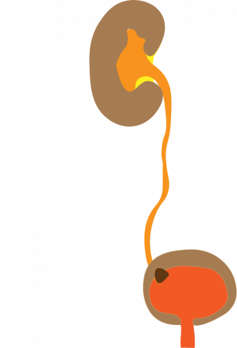
| Renal colic | - Hydronephrosis
- Renal stone with acoustic shadowing – they do not always produce acoustic shadowing
- Twinkling artifact
- Asymmetric ureteric jets
|
Leave A Comment